


Expand Your Knowledge
Our resource center archives our case studies, published articles, blogs, webinars, and image galleries. Discover ways microscopy has made a meaningful impact.
Microparticles may occur as unwanted visitors, but they can also be important components of many different types of products. For example, a manufacturer may produce a powdered product that must adhere to a specific particle size distribution range.
At MVA Scientific Consultants, we have the capability to go beyond routine light scattering methods of particle sizing. Using either of our scanning electron microscopes (SEMs), we can perform automated particle analysis to determine the size and shape of tens of thousands of individual particles in a single sample.
The first step in an SEM automated particle analysis is to acquire images with sufficient contrast between the background and the particles so that an image analysis algorithm is capable of differentiating between them (Figure 1). For automated image analysis systems, a “particle” is defined as a set of contiguous pixels all of which are brighter (or more rarely, darker) than the threshold brightness used to define the surrounding “background” pixels.
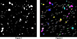
The next step is the recognition of particles by the analysis system, which is part of the SEM software. Figure 2 shows the same field of view as Figure 1, now indicating all of the particles that the system has found. The analysis system saves the location of each particle and determines the two-dimensional size and shape parameters for each one. Typical parameters include maximum, minimum and average diameters, perimeter and aspect ratio.
When every particle in the field of view is recognized and its dimensions saved, the microscope moves to a new field of view and the process is repeated until a set number of particles or a predetermined number of fields of view have been analyzed. In this way tens of thousands of particles can be characterized in terms of size and shape in a few hours without operator involvement beyond the initial setup.
The final results are tabulated, giving a complete picture of the particle sizes and shapes. This tabulation is entirely customizable since all of the data is stored for each individual particle.
If you think your facility or clients could benefit from automated particle sizing analysis, please don’t hesitate to contact us.